
1 The structure of DNA
(a) Structure of DNA —nucleotides (deoxyribose sugar, phosphate and base), sugar–phosphate backbone, base pairing (adenine – thymine and guanine – cytosine), by hydrogen bonds and double stranded antiparallel structure, with deoxyribose and phosphate at 3′ and 5′ ends of each strand respectively, forming a double helix.
Revision Resources:
 |
 |
 |
|
|
All cells, bacterial (prokaryotic) or more complex cells such as animal, plant or fungal cells (eukaryotic) store their genetic information in the form of DNA. Many viruses, which are not made of cells also use DNA, although some carry genetic information as RNA. It is the DNA base sequence (order of the bases) which ultimately determines the phenotype of an organism.
From your National 5 studies, you will know something of the structure of DNA, and should be aware of the way in which this information is held in the molecule. At Higher level, a more detailed understanding of the structure of the DNA molecule and its replication is required. In addition an understanding of the form in which DNA is stored is included. First a review of the basics.
DNA Structure:
The following video will help you understand this area. (note at time 3:11, where the arrangement of the polymer is being shown, the authors have incorrectly labelled the 3′ carbon on this section)
DNA stands for DeoxyriboNucleic Acid). A DNA strand is made up of tiny units called nucleotides (Figure 1), each consisting of a deoxyribose sugar, base and phosphate.

Figure 1: A DNA nucleotide
The deoxyribose sugar is a pentagon (5-sided), and each carbon in the pentagon is given a number. Examine the diagram below (Figure 2) The pentagon is closed at the top by an oxygen which isn’t numbered; carbon number 5 sits out of the pentagon ring, attached on to carbon number 4. The base of the nucleotide is attached at carbon number 1 and carbon number 3 (3’) contains an “OH” group.
As you can see in the diagram below, the phosphate group is attached to carbon number 5. (5’).

Figure 2: Detailed structure of a nucleotide, showing the numbering and arrangement of the carbon atoms in the deoxyribose sugar ring.
DNA nucleotides can be joined together by attaching them between the phosphate group (5’) and the OH group (3’). The bond formed is a strong, covalent bond and is known as a sugar phosphate bond (Figure 3). With many DNA nucleotides joined together a sugar-phosphate backbone is formed.

Figure 3: Formation of the sugar phosphate backbone
Many nucleotides are joined together to form a DNA strand. Two DNA strands then wind around each other to form a DNA molecule. The shape of the molecule is known as a double helix. (Figure 4). If you examine the diagram (Figure 4) closely you will see that the strands on either side of the DNA molecule run in opposite directions. In the diagram, the left hand strand has a phosphate group uppermost (hence carbon 5’), and the right hand strand has the other end uppermost (carbon number 3, which contains an OH, 3’) remind yourself of 3’ and 5’ using the diagram above (Figure 2, page 3). With the strands running in opposite directions in this way, the DNA molecule is said to be antiparallel.

Figure 4: Formation of the DNA strand and double helix conformation (shape).
The two strands are held together by weak hydrogen bonds between the bases. This means they are easy to break apart, an important feature for DNA replication (). The bases will only join up according to DNA base-pairing rules:
The bases which pair up on opposite strands are said to be complementary with:-
- § Adenine always pairing with Thymine
- § Cytosine always pairing with Guanine
Gel electrophoresis is a technique used to spearate DNA strands of different lengths. It utilises the fact that DNA is a negatively charged molecule. The DNA is placed in small wells at the top of an agarose gel, and an electric field applied. The current is switched on and the DNA is attracted to the positive electrode (anode). The longer the piece of DNA, the harder it is for it to move through the gel – so the larger fragments are found nearer the wells, whilst the smaller fragments move faster and are located nearer the positive electrode (anode) at the opposite side from the wells.
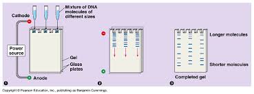
The DNA must be stained with before it can be visualised in the gel.
The following links and websites will help provide background to your understanding of DNA:
| Chargaff’s Experiments | Griffith’s and Avery’s Experiments | Hershey & Chase Experiments |